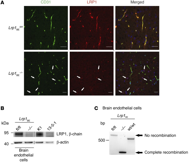
Figure 1. Full deletion of Lrp1 in Lrp1BE–/– mice.
(A) Immunofluorescent staining for endothelial marker CD31 and LRP1 in cortical brain sections demonstrated complete knockout of Lrp1 in brain endothelium of Lrp1BE–/– animals, while Lrp1 expression in surrounding cells remained unaffected (white arrows). DRAQ5 was used to stain cell nuclei. Scale bar: 20 μm. (B) Immunostaining in isolated endothelial cells showed knockout of Lrp1 in Lrp1BE–/– mice. Primary cortical endothelial cell and control lysates of LRP1-expressing CHO cells (K1) and LRP1 knockout (13‑5-1) cells were analyzed on the same Western blot but rearranged for clearer presentation. An anti–β-actin immunoblot is shown as a loading control. (C) PCR analysis revealed complete Cre-mediated excision of the loxP-flanked Lrp1 allele in brain endothelium. Endothelial genomic DNA was used for PCR detecting the WT (WT/WT, 507 bp), the loxP-flanked (fl/fl, 541 bp), and the excised allele (–/–, 325 bp) simultaneously in one reaction. Data show representative results from experiments performed in triplicate.

