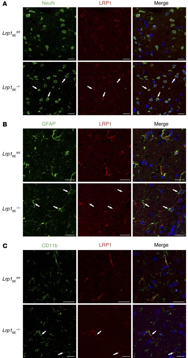
Figure 3. Brain endothelial–specific deletion of Lrp1 in Lrp1BE–/– mice.
Immunofluorescent staining in cortical brain sections for LRP1 and (A) NeuN-positive neuronal cells, (B) GFAP-positive astrocytes, and (C) CD11b-positive microglia and macrophages to determine potential recombination in macrophages/microglia, neurons, and astrocytes revealed no differences between genotypes. Scale bar: 20 μm. Data show representative results from experiments performed in triplicate.

