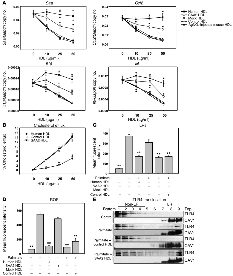
Figure 3. SAA-enriched HDL partly loses its antiinflammatory effect on adipocytes.
(A–E) 3T3-L1 adipocytes were preexposed for 6 hours to human HDL or human HDL isolated after exposure to lentiviral-transduced 293 HEK cells, as described in Methods. HDL concentration was as indicated (A and B) or 50 μg protein/ml (C and D). Thereafter, adipocytes were treated as described in the legend to Figure 1. Saa3, Ccl2, Il1β, and Il6 gene expression (A); cholesterol efflux to human, control, and SAA2-HDL (B); LR content (C); ROS generation (D); and TLR4 translocation to LRs (E). An antibody to caveolin 1 (CAV1) was used to stain LRs. Fractions 7–9 contain LRs, and fractions 1–4 are non–LR-containing fractions. Data represent mean ± SD. Data are representative of at least 3 independent experiments. *P < 0.001 vs. control-HDL, **P < 0.001 vs. SAA2-HDL. ANOVA and Bonferroni post-hoc test.

