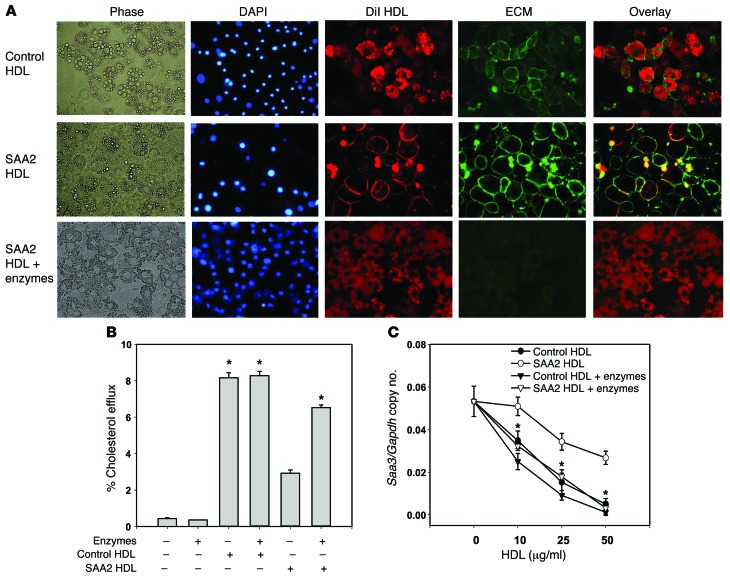
Figure 6. SAA-enriched HDL colocalizes with the adipocyte cell surface.
(A) Control HDL, which was reisolated from conditioned media from cells not transduced with lentivirus, and SAA2-HDL, which was reisolated from conditioned media from cells transduced with a lentivirus-encoding mouse SAA2.1 protein were labeled with DiI (red). 3T3-L1 adipocytes were exposed to these HDL preparations and stained as described in the legend to Figure 4. Original magnification, ×400. SAA2-HDL colocalized with cell surface–associated ECM staining. Pretreatment of adipocytes with chondroitin ABC lyase and heparitinase prior to exposure of SAA2-HDL resulted in a similar distribution of dye to that seen with control HDL. (B and C) Representative fluorescence images of 3 independent experiments are shown. After predigestion of the proteoglycans of 3T3-L1 adipocytes with chondroitin ABC lyase and heparitinase for 1 hour, exposure to SAA2-HDL (50 μg protein/ml) for 6 hours resulted in a restoration of cholesterol efflux (B) and Saa3 gene expression (C) toward that seen with control HDL. Data represent mean ± SD. Data are representative of at least 3 independent experiments. *P < 0.001 vs. SAA2-HDL. ANOVA and Bonferroni post-hoc test.

