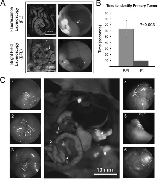
Figure 6.

Enhanced visualization of primary and metastatic pancreatic cancer through fluorescence laparoscopy. (A) During laparoscopy, malignancies were easily visualized using the fluorescence mode (FL) with a fluorescent-labeled antibody. The visualization of tumors using the bright field (BFL) mode was hindered, in comparison to FL. (B) Time to identify the primary tumor using FL and BFL showed that FL was a much faster technique. (C) Using FL, both primary and metastatic lesions were easily visualized in each case. The center image represents shows six tumors in the abdomen labeled 1–6. The corresponding images of both primary tumors (4 and 5) and metastatic disease (1, 2, and 3) are shown individually. Reprinted with permission from ref (131). Copyright 2012 H.G.E. Update Medical Publishing Athens.
