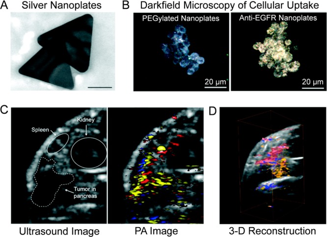
Figure 7.

Photoacoustic imaging of pancreatic cancer using antibody-targeted silver nanoplates. (A) The edge lengths of silver nanoplates were 218 ± 35.6 nm. (B) Darkfield microscopy showed increased cellular uptake of antibody-modified nanoplates (left) in comparison to PEGylated nanoplates (right). (C) Two-dimensional cross sections of orthotopic tumors allowed for delineation of organs and produced a photoacoustic signal from antibody-modified silver nanoplates (yellow), oxygenated blood (red), and deoxygenated blood (blue). (D) Image reconstruction produced a 3-dimensional representation of orthotopic pancreatic tumor model with the photoacoustic signal. Reprinted with permission from ref (169). Copyright 2012 American Chemical Society.
