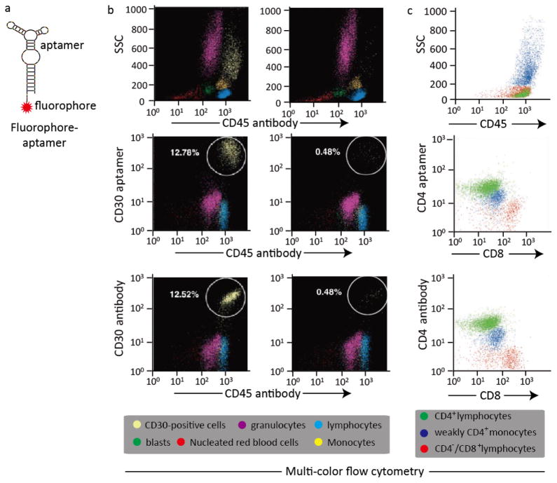
Fig. 1. Aptamer-based fluorescent probe for multi-color flow cytometric analysis.
(a) Design of a simple, sensitive and versatile fluorophore-aptamer probe for cancer cell detection or in vivo tumor imaging; (b) Multi-color flow cytometric analysis of lymphoma cells by aptamer probes and antibodies. CD30-positive lymphoma cells were mixed with fresh normal marrow cells and stained with AmCyan-labeled CD45 antibodies, and Cy5-labeled CD30 aptamers simultaneously, or FITC-labeled CD30 antibodies as standard control. The individual cellular populations in the cell mixture including nucleated red blood cells, blasts, lymphocytes, granulocytes, monocytes and CD30-positive lymphoma cells, were separated and gated according to the side scatter (SSC) and CD45 panels. Multi-color flow cytometric analysis revealed that both aptamer probes and antibodies detected the same population of CD30-positive lymphoma cells with identical specificities and sensitivities; (c) Cells from patients’ pleural fluids were collected and incubated with different fluorophore-labeled CD4 aptamers (CD4 antibodies as standard control), CD8 and CD45 antibody simultaneously. Multi-color flow cytometric analysis revealed that the CD4 aptamers also showed similar staining patterns and numbers for the as-prepared cells as the CD4 antibodies.

