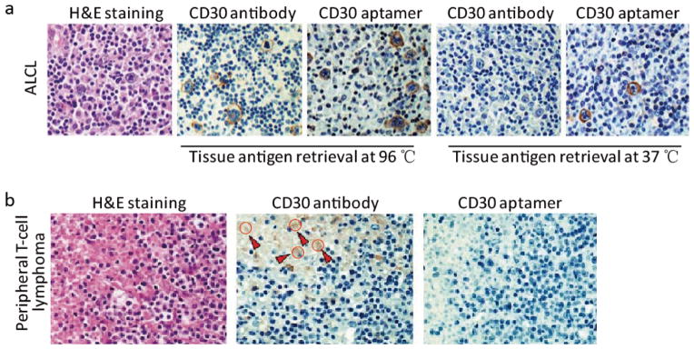
Fig. 3. Aptamer probes for IHC staining of FFPE tumor tissues.
(a) Tissue sections of CD30-positive anaplastic large cell lymphoma (ALCL) were immunostained with aptamer probes, or antibodies as standard control. After antigen retrieval at 37 °C and probing for 20 min, tissue sections were probed with aptamers and lymphoma cells were specifically immunostained. In contrast, antibody immunostaining of lymphoma cells required higher antigen retrieval temperature (97 °C) for a long probing time (90 min); (b) Aptamer probes showed non-specific staining (brown color) in the tumor necrotic area compared to the antibody stain (depicted with red arrows and circles).

