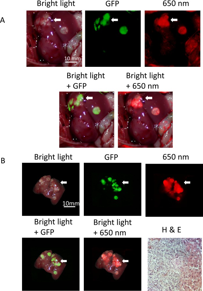
Fig 4. Imaging of IGF-1R targeting of liver metastasis.
Anti-IGF-1R conjugated to PEGylated 650 nm dye selectively labeled the HCT 116-GFP metastatic tumors. Both in vivo (A) and ex vivo (B) imaging show that the 650 nm fluorophore-conjugated IGF-1R antibodies co-localized with HCT 116-GFP fluorescence and more accurately demarked the tumor compared to bright light imaging (white arrowheads). H & E staining of tissue sample (x200) expressing fluorescence confirms the presence of metastatic tumor in the mouse liver.

