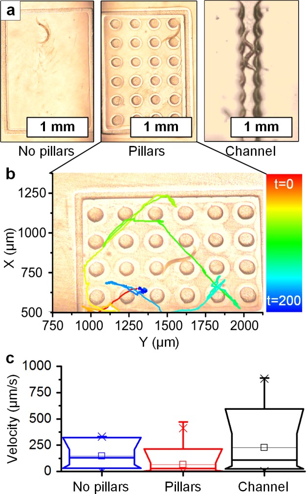
Fig 3. In situ photopolymerization of microstructures resulting in physical confinement of a C. elegans.

(a) Three exemplary assays built sequentially (from left to right): an open frame, an array of micropillars (100 μm diameter), and a rippled microchannel (approx. 200 μm wide). (b) Tracking of the worm motion over a time period of 200s, within the pillar array. (c) Box-whisker plots of velocity in each configuration, showing that sequential confinement increases the maximum velocity at which the worm pushes against the surface of features. S1 Video shows the experiment.
