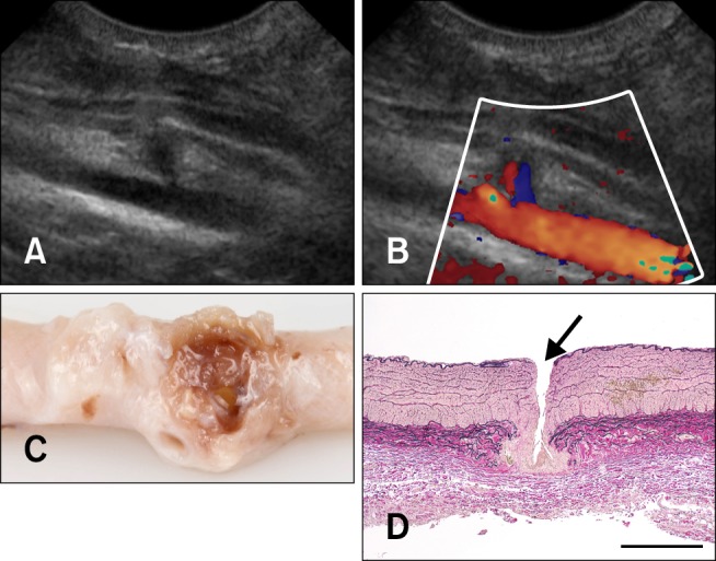
Fig. 5. The femoral artery of a Boerboel dog 6 days after removal of the introducer-sheath. (A) 2-dimensional ultrasonography shows a small local aneurysm at the site of the puncture. (B) Color Doppler ultrasonography shows a patent lumen (the same longitudinal image as on panel A). (C) The dissected femoral artery (stored in formalin). Granulation tissue can be seen around the puncture site. (D) Photomicrogram of the femoral artery around the puncture site (arrow); longitudinal section. Lawson stain, scale bar = 500 µm.

