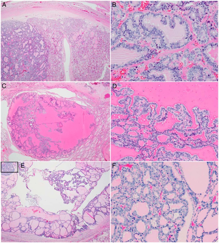
Figure 1.
Pathological features of papillary carcinoma arising in follicular nodules in three siblings with germline DICER1 mutations. A, Low-power view of a 1.3-cm encapsulated follicular nodule in patient B. Note the apparent clonal outgrowth in the left third of the photomicrograph. B, High-power view of this area shows cytological features of papillary carcinoma. C, Low-power view of a section from a 1.6-cm partially encapsulated cystic nodule from patient A. D, Medium-power view shows papillae with overlapping nuclei, nuclear clearing, grooves, and rare pseudoinclusions. E, The brother of these two siblings had two small foci of papillary carcinoma within larger encapsulated follicular nodules with cystic change and papillary hyperplasia. A low-power view of a left lobe follicular nodule with small focus of papillary carcinoma (box) is shown in panel E. F, High-power view of papillary carcinoma shown in panel E.

