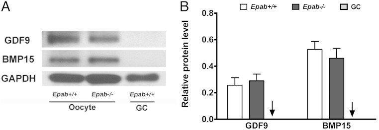
Figure 3.
The expression of GDF9 and BMP15 is not altered in the oocytes of Epab−/− mice. A, GDF9 and BMP15 expression was detected by Western blot analysis using 300 GV oocytes from WT or Epab−/− mice. GCs from WT mice were used as a negative control. B, Band intensities were analyzed using densitometry and normalized to GAPDH. Data are represented as the mean ± SEM from 3 independent experiments. There was no significant difference in GDF9 or BMP15 expression between WT and Epab−/− oocytes. Arrow designates WT GC bar.

