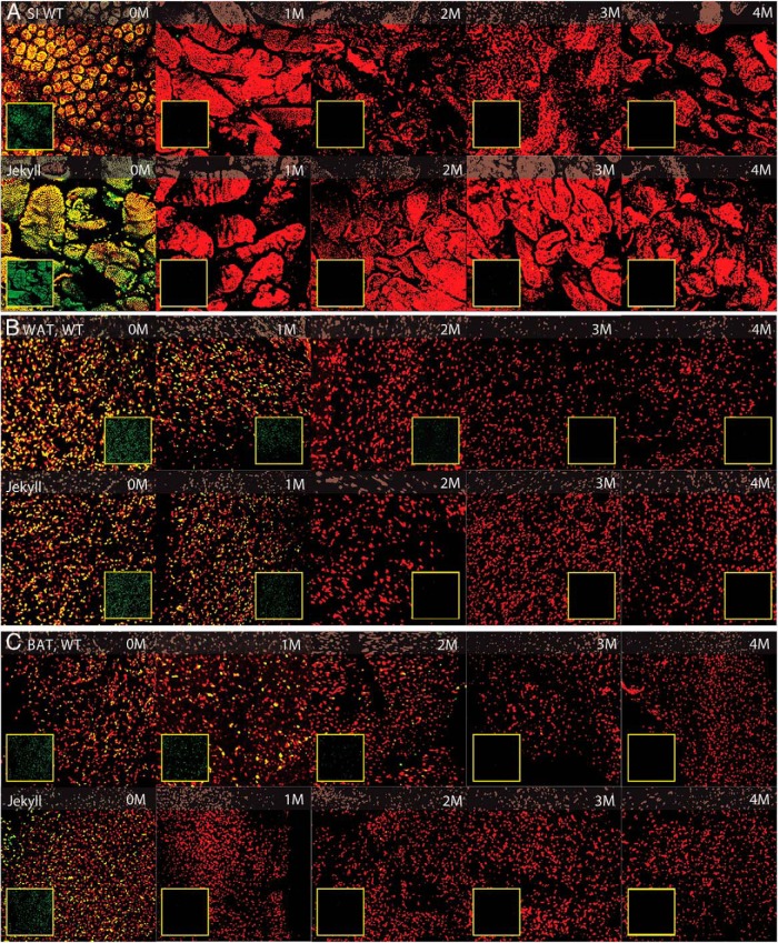
Figure 2.
Histological assessment of adipose tissue and small intestine (SI). Wild-type (WT) and Jekyll H2B-GFPxM2 mice were pulsed with doxycycline for 2 weeks followed by 0-, 1-, 2-, 3-, and 4-month chase periods. Whole-mount SI and WAT and BAT were imaged by confocal microscopy at ×250 (red, propidium iodide-stained nuclei; green, LRC nuclei [inset boxes]; yellow, overlay). Representative composite image stacks representing 25–4-μm digital sections are shown for each chase period.

