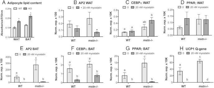
Figure 6.
Differentiation of adipose stromal vascular cells. A, WAT and BAT cells from wild-type and mstn−/− (KO) mice were differentiated and adipocyte lipid content, as a marker of terminal differentiation, was quantified with Oil Red O staining (n = 6/group; *, P ≤ .05 compared with wild type [WT]). Cells were also treated with or without recombinant myostatin (A–H), and gene expression of adipogenic markers was quantified. Markers include adipocyte lipid-binding protein (AP2), C/EBPα, and PPARγ, which are common to both WAT and BAT, as well as uncoupling protein (UCP)1, which is unique to BAT. In C–H, significant differences among all groups are indicated by different letters (n = 6/group, P ≤ .05), and in B by an asterisks (compared with 0 control).

