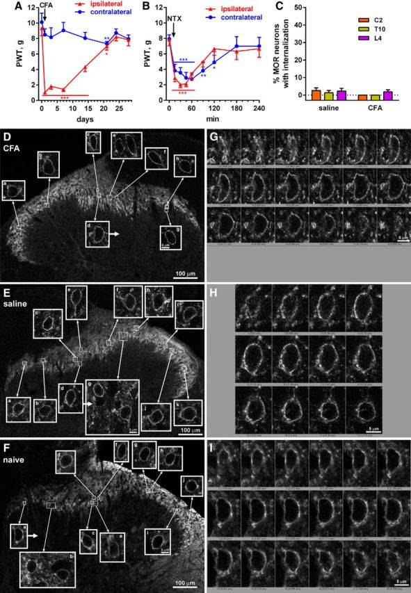
Figure 7.

Absence of MOR internalization in the spinal cord during the remission phase of latent sensitization. A, Rats (n = 6) implanted with intrathecal catheters were injected in the hindpaw with 50 μl of CFA subcutaneously and responses to von Frey hairs were tested. B, On day 28, the rats received NTX (1 mg/kg, s.c.) and PWTs were tested. Holm-Sidak's post hoc tests: *p < 0.05, **p < 0.01, ***p < 0.001 compared with baseline (time 0 d or 0 min). C, On day 30, rats injected with CFA (n = 4) or saline-injected controls (n = 4) received intrathecal injections of peptidase inhibitors (amastatin, phosphoramidon, and captopril, 100 nmol) and fixed 15 min later. MOR immunohistochemistry was performed in spinal segments C2, T10, and L4 to measure MOR internalization. D, Images taken from the L4 segment of a rat 30 d after injecting CFA in the hindpaw; MORs are not internalized. E, Images taken from the L4 segment of a rat 30 d after injecting saline in the hindpaw; MORs are not internalized. F, Images taken from the L4 segment of a naive rat; MORs are not internalized. Images are single optical sections taken with a 10× objective (main panels, voxel size 830 × 830 × 5983 nm, scale bar 100 μm) or a 63× objective (insets, voxel size 132 × 132 × 383 nm, scale bar 5 μm). G, Confocal stack of cell d (arrow) in D. H, Confocal stack of cell d (arrow) in E. I, Confocal stack of cell a (arrow) in F.
