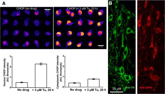
Figure 1.
Validation of antibodies. A, CHOP antibody staining in WT mouse ventral midbrain cultured cells that are also positive for TH (data not shown). Left, No drug. Right, Twenty hours in 3 μm Tu. The left and right graphs show the nuclear and cytoplasmic CHOP intensity, respectively. B, ATF6 antibody staining in slices from adult mouse substantia nigra. Double staining with anti-TH and anti-ATF6 shows that all DA neurons also stain for ATF6.

