
Figure 7.
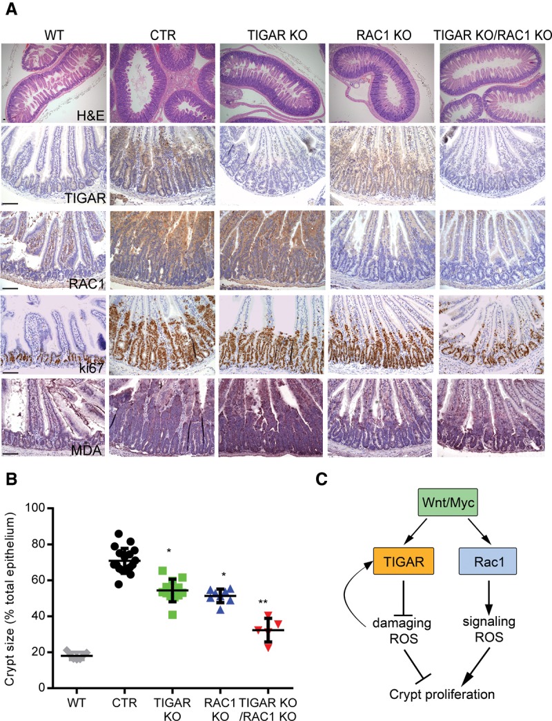
In vivo deletion of both TIGAR and RAC1 can synergistically decrease hyperproliferation of Apc-deficient intestinal crypts. (A) H&E staining (top row), TIGAR staining (second row), RAC1 staining (third row), Ki67 staining (fourth row), and MDA staining (bottom row) of small intestines from wild-type (WT), Ahcre+Apcfl/fl (CTR), Ahcre+Apcfl/flTIGARfl/fl (TIGAR knockout [KO]), Ahcre+Apcfl/flRac1fl/fl (RAC1 knockout), and Ahcre+Apcfl/flTIGARfl/flRac1fl/fl (TIGAR/RAC1 knockout) animals 3 d after β-napthoflavone induction of Ahcre. Bars, 100 µm. (B) Measurements of crypt thickness from A as a percentage of total thickness of epithelium. (*) P < 0.05 compared with CTR; (**) P < 0.05 compared with TIGAR knockout or RAC1 knockout. CTR, n = 18; TIGAR, n = 12, RAC1 knockout, n = 7; TIGAR knockout RAC1 knockout, n = 5. (C) Proposed model of the role of ROS after Wnt signaling activation.