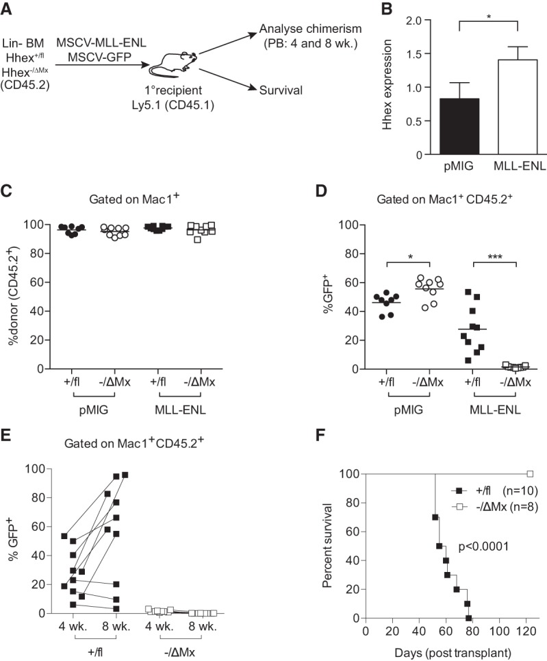
Figure 2.

Hhex is required for initiation of AML by MLL-ENL. (A) Schematic diagram of experimental design using Hhex+/fl and Hhex−/ΔMx mice. (B) Hhex expression in virally transduced BM cells at 2 d after transduction as assessed by quantitative PCR. (C–E) Peripheral blood analysis of recipient mice injected with MSCV-IRES-GFP (MIG) and MIG-MLL-ENL transduced lineage-depleted BM from mice of the indicated Hhex genotypes. (C) Percentage of myeloid (Mac1+) cells that are donor-derived (CD45.2+) at 4 wk after transplant. (D) Percentage of donor-derived myeloid cells (Mac1+CD45.2+) that are virally transduced (GFP+) at 4 wk after transplant. Lines show the mean. (E) As in D, showing 4- and 8-wk time points of MLL-ENL transduced BM recipients of the indicated Hhex genotypes. Lines connect sequential samples taken from individual mice. (F) Kaplan-Meier survival curve of recipients of MLL-ENL transduced BM of the indicated Hhex genotypes (analyzed in C–E). Data in C–F are a combination of two separate experiments. (*) P < 0.05; (***) P < 0.001, Student's t-test. In F, P was determined using a log rank (Mantel-Cox) test.
