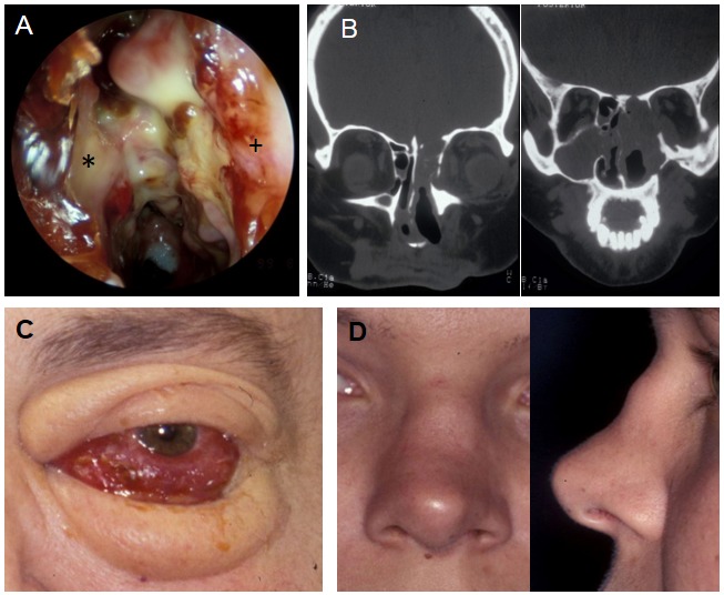
Figure 3. A) Nasal cavity in GPA with granulations, destruction, bacterial infection, left nasal cavity (choanal #, nasal septum*, lateral nasal wall+), B) CT scan of the same patient, destruction of the orbital wall and frontal skull base, C) The same patient, clinical aspect, D) Saddle nose deformity in GPA.

