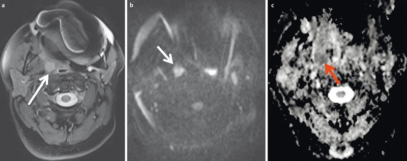
Figure 27. 49-year-old patient after resection of a lip carcinoma on the right side.

a) Axial T2w images demonstrate a solitary retropharyngeal lymph node (arrow). Significant artifacts due to metallic dental implants.
b) The lymph node (arrow) appears hyperintense on diffusion-weighted images.
c) The lymph node (arrow) appears hypointense on the ADC map. Calculated ADC was 0.65×10⁻³ mm2/s, indicating malignancy. This was confirmed by histology.
