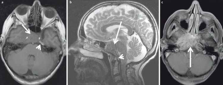
Figure 30. 53-year-old patient with squamous cell carcinoma of the nasal cavity and osseous infiltration of the skull base.

a) On T1w images the hypointense tumor (arrow) replaces the hyperintense fatty marrow of the skull base (arrowhead).
b) On sagittal T2w images (arrow) the tumor also appears hypointense compared to bone marrow (arrowhead).
c) There is avid enhancement after contrast administration.
