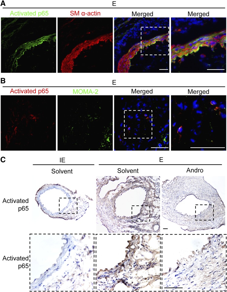
Fig. 2.
Andro attenuates NF-κB activation in aortic tissues. (A and B) Representative images of arterial sections coimmunostained for activated p65 (green) and smooth muscle α-actin (smooth muscle α-actin; red) (A) or activated p65 (red) and MOMA-2 )green) (B) overlaid with 4′,6-diamidino-2-phenylindole (blue). Areas highlighted by white dashed boxes are shown at higher magnification on the right. Arteries were harvested 7 days after elastase perfusion. Scale bar = 50 μm. (C) Representative images of arterial sections immunohistochemically stained for activated p65. E, elastase; IE, inactivated elastase. Areas highlighted by black dashed boxes are shown at higher magnification at the bottom. Arteries were harvested 14 days after elastase perfusion. Scale bar = 100 μm.

