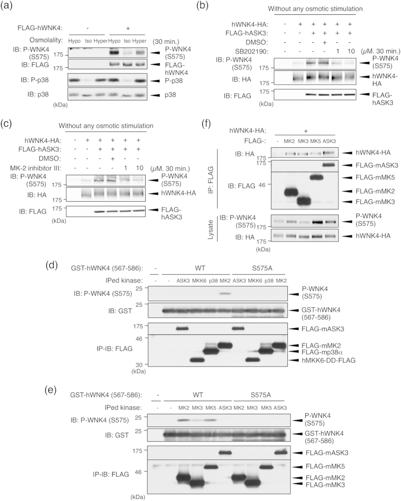
Figure 3. ASK3-dependent WNK4 Ser575 phosphorylation is mediated by the p38MAPK-MK pathway.
(a) WNK4 Ser575 phosphorylation is induced by the osmotic stress. HEK293A cells were transfected with FLAG-tagged human WNK4 (FLAG-hWNK4). At 48 h after transfection, cells were stimulated with indicated stimulation buffer for 30 minutes. Full-length blots are presented in Supplementary Fig. S7a. (b) Ser575 phosphorylation induced by ASK3 co-expression without any osmotic stress is attenuated by p38MAPK inhibition. HEK293A cells were transiently transfected with hWNK4-HA and FLAG-hASK3. Before lysis, the cells were treated with DMSO or SB202190 at the indicated concentrations for 30 min. Full-length blots are presented in Supplementary Fig. S7b. (c) Ser575 phosphorylation induced by ASK3 co-expression without any osmotic stress is attenuated by MK2 inhibition. HEK293A cells were transiently transfected with hWNK4-HA and FLAG-hASK3. Before lysis, the cells were treated with DMSO or MK-2 inhibitor III at the indicated concentrations for 30 min. Full-length blots are presented in Supplementary Fig. S7b. (d) Only MK2 directly phosphorylates WNK4 Ser575 among the components of the ASK3-MKK6-p38MAPK-MK pathway. mIMCD3 cells were transiently transfected with FLAG-tagged mouse ASK3, a constitutively active human MKK6 mutant (MKK6-DD), mouse p38α and mouse MK2. Immunoprecipitated kinases were incubated with GST-hWNK4 (567–586) and Mg-ATP for 15 min at 30 °C. Full-length blots are presented in Supplementary Fig. S7c. (e) MK2, MK3 and MK5 are capable of directly phosphorylating WNK4 Ser575. mIMCD3 cells were transiently transfected with FLAG-tagged mouse MK2, mouse MK3 or mouse MK5. Immunoprecipitated kinases were incubated with GST-hWNK4 (567–586) and Mg-ATP for 15 min at 30 °C. Full-length blots are presented in Supplementary Fig. S7d. (f) Interaction between WNK4 and MK kinases. mIMCD3 cells were transiently transfected with hWNK4-HA and FLAG-tagged mouse MK2, MK3 and MK5. Cells were lysed at 24 h after transfection, and FLAG-tagged MK kinases were immunoprecipitated with anti-FLAG antibody beads. Full-length blots are presented in Supplementary Fig. S8.

