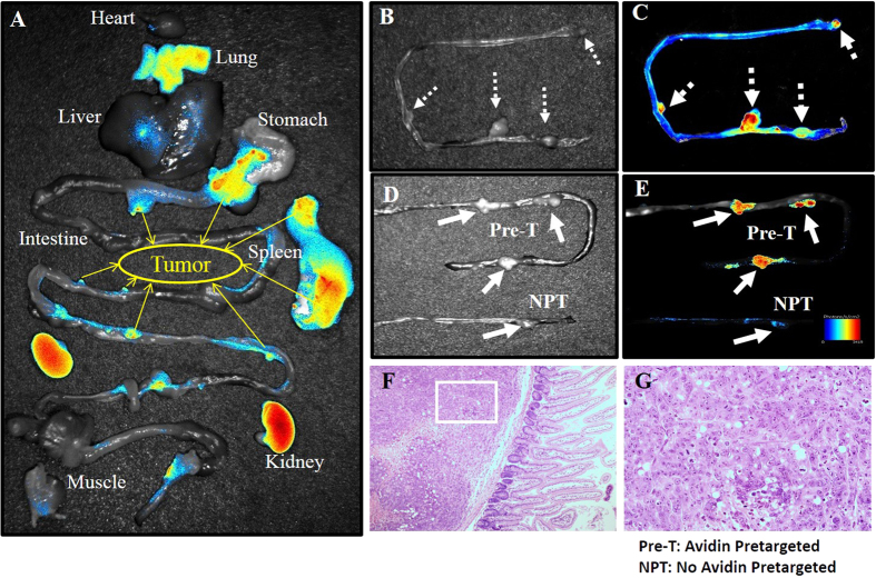
Figure 3. NIR-fluorescence imaging of 99mTc-HYNIC-lys(Cy5.5)-PEG4-biotin in nude mice bearing LS180 colon tumors.
(A) Tissue uptake of HYNIC-lys(Cy5.5)-PEG4-biotin. Yellow arrows mark tumors. (B,C) Local NIRF imaging of mouse intestines with colon tumors. Dashed arrows mark tumors. (D,E) Contrast between tumor uptakes of HYNIC-lys(Cy5.5)-PEG4-biotin with and without pretargeted avidin. Solid line arrows mark the tumors. (F,G) HE staining of selected tumor section.

