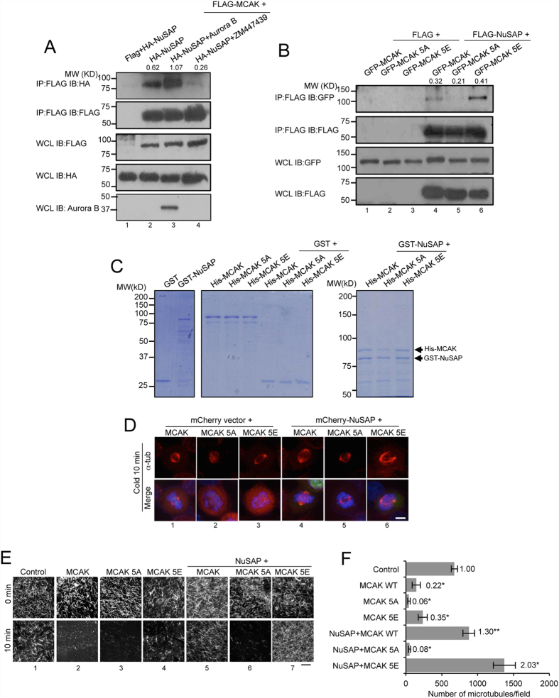
Figure 4. Aurora B positively regulates the function of NuSAP on MCAK depolymerisation activity.
(A) Aurora B enhances the binding between NuSAP and MCAK. Whole cell lysates of 293T cells co-transfected with FLAG vector or FLAG-MCAK and HA-NuSAP in the presence of either Aurora B or 2 μM ZM447439 for 45 min were collected for co-immunoprecipitation (Co-IP) using FLAG-M2 beads. Immunoprecipitated proteins and whole cell lysates were detected with anti-HA and anti-FLAG antibodies. Quantification of the gel images with Image J indicates that the ratio of immunoprecipitated NuSAP to MCAK. (B) Whole cell lysates of HEK 293T cells co-transfected with a FLAG vector or FLAG-NuSAP and GFP-MCAK, GFP-MCAK 5A, or GFP-MCAK 5E were collected for Co-IP using FLAG M2-beads. Immunoprecipitated proteins and whole cell lysates were detected with anti-GFP and anti-FLAG antibodies. Quantification of the gel images with Image J indicates that the ratio of immunoprecipitated MCAK to NuSAP. (C) MCAK and MCAK 5E bind NuSAP in vitro. Purified His-MCAK, MCAK 5A, or MCAK 5E proteins were incubated with GST or GST-NuSAP and detected with Coomassie blue staining and western blotting in 10% (left panels) and 6% (right panel) SDS-PAGE. (D) Kinetochore microtubules in cold-treated metaphase HeLa cells expressing GFP-MCAK, GFP-MCAK 5A, or GFP-MCAK 5E with mCherry vector or mCherry-NuSAP. Cells were stained with anti-α-tubulin antibody and Hoechst 333342. Scale bar, 5 μm. (E) Microtubule depolymerisation assays with MCAK and NuSAP. NuSAP (100 nM) and 20 nM MCAK WT, MCAK 5A, or MCAK 5E proteins were incubated with 1.5 μM microtubules at 37 °C for 10 min. Scale bar, 20 μm. (F) Bar chart representing the number of microtubules in control, MCAK-, MCAK 5A-, and MCAK 5E-treated samples with or without NuSAP protein after a 10 min incubation. Three independent experiments were conducted. Error bars represent ± SD. *p < 0.001.

