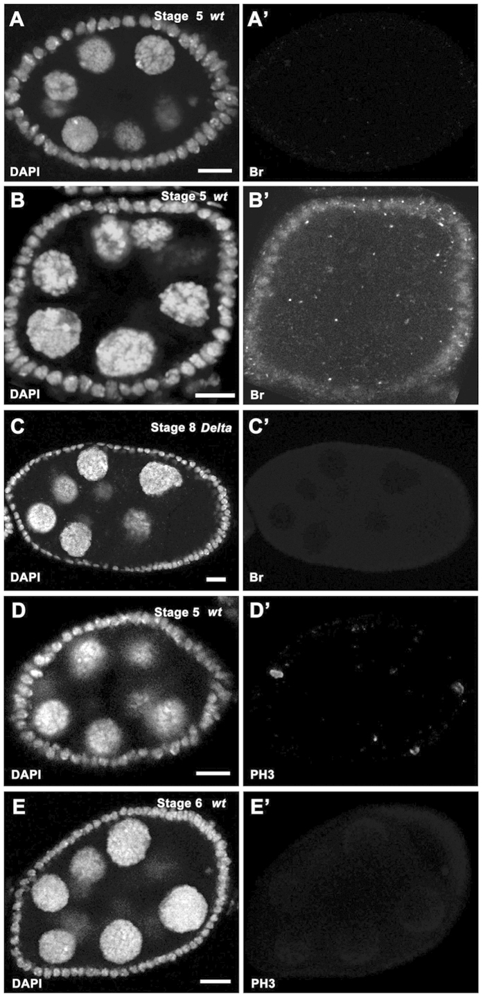
Figure 7. Experimental applications of stage identification.
In all panels, DAPI staining (white in A–E) marks cell nuclei. (A-A’) Br expression (white in A’) was absent in a stage-5 wildtype egg chamber (egg chamber size: 2273.6 μm2). (B-B’) Br expression (white in B’) was detected in a stage-5 wildtype egg chamber (egg chamber size: 2175.4 μm2). (C-C’) Follicle cells covering the Dlrev10 germline clone in a stage-8 egg chamber (egg chamber size: 7908 μm2) did not show detectable Br expression (white in C’). (D-D’) Mitotic marker PH3 staining (white in D’) was detected in follicle cells of a stage-5 egg chamber (egg chamber size: 2999.4 μm2). (E-E’) Mitotic marker PH3 staining (white in E’) was absent in follicle cells of a stage-6 egg chamber (egg chamber size: 4606.1 μm2). Anterior is to the left. Bars, 10 μm.

