
Figure 7. PDE1 inhibitor IC86340 induces PDGFRβ protein degradation via endocytosis.
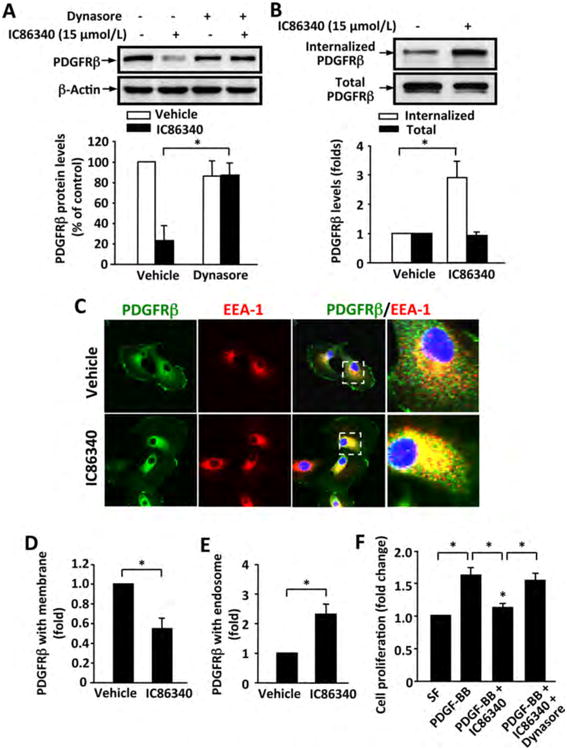
(A) Dynamin inhibitor dynasore blocked IC86340-induced PDGFRβ protein reduction. Rat aortic SMCs were treated with 15 μmol/L IC86340 with or without 2.5 μmol/L dynasore for 24 h in DMEM containing 0.1% FBS. (B) IC86340 induced PDGFRβ internalization. Rat aortic SMC membrane proteins were labeled with biotin and stimulated with 15 μmol/L IC86340 for 12 h in DMEM containing 0.1% FBS. Cell lysates were immunoprecipitated with streptavidin beads, and the recovered internalized biotinylated PDGFRβ were immunoblotted with PDGFRβ antibody. (C-E) IC86340 increased PDGFRβ co-localization with endosomes. Rat aortic SMCs were treated with 15 μmol/L IC86340 for 12 h in DMEM containing 0.1% FBS. PDGFRβ and early endosome marker EEA-1 were determined by immunostaining. C, representative images showing the localization of PDGFRβ in plasma membrane and endosomes. Selected boxes were enlarged for better view. D, quantitative data of PDGFRβ on cell membrane. E, quantitative data of PDGFRβ in endosomes. (F) Dynasore abrogated the inhibitory effect of IC86340 on SMC growth. Rat aortic SMCs were treated with IC86340, dynasore or a combination of IC86340 and dynasore, and stimulated with 50 ng/ml PDGF-BB for 48 h. Cell proliferation was measured by SRB assay. Values are mean ± SD of (n=3). *P < 0.05.