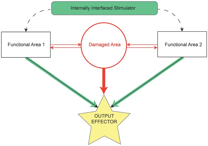
Figure 3.

Schematic by which internally contained stimulation devices restore lost function resulting from damaged or missing tissue. Before damage, the area of interest (red circle) and functionally related areas (rectangles) relay information between each other and effectors (solid arrows) of some output task (yellow star). The majority of information in controlling task output initially comes from the damaged area (thick red arrow), but may also arrive, although to a lesser extent, from functionally related areas (thin black arrows). Following injury, connections to and from the damaged area are lost (all red elements). The stimulation device serves as a direct bridge between functional areas, allowing strengthened output (thick green arrows) from those areas to the output effectors and thereby restoring some degree of lost functionality.
