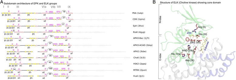
Fig. 1.

The kinase core domain in a representative set of EPKs and ELKs. a. Secondary structure labels are indicated below the sequence, and Hanks and Hunter subdomain notations are shown above the sequence. Insert segments longer than 5 residues are indicated as arcs in the sequence, and the numbers indicate the average insert size. b. Core domain highlighted in the crystal structure of choline kinase
