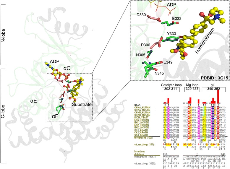
Fig. 4.

The substrate recognition region in Choline kinases. On the left is shown the structural context of the substrate binding region. The inset shows conserved residues within the substrate binding region that are most distinctive of these kinases based on mcBPPS analysis. In the structure figures, the green region corresponds to the core domain whereas the black regions are outside of the core. Residues that are Choline kinase-specific are shown with green carbon atoms and catalytic residues are shown in light pink carbon atoms. The substrate analog hemicolinium is shown in yellow CPK representation. A Contrast Hierarchical Alignment (CHA) in the bottom right panel shows the constraints imposed on residues in key regions of the kinase domain. CHA shows representative Choline kinase sequences as the display alignment; all Choline kinase sequences (702 sequences) as foreground and other ELK sequences (15283 sequences) as background. CHA coloring scheme and representation is similar to that described in Fig. 3d
