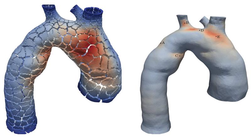
Figure 4.
Mesh of an aorta seen from above showing the brachiocephalic artery, and the left common carotid and subclavian arteries. The fine mesh consists of 5 418 594 tetrahedrons and 1 055 901 vertices, while colors indicate the displacement field with an internal pressure of 1 mmHg. Additionally, the splits show the decomposition of the mesh into 480 subdomains (left). Coarser mesh consisting of 720 060 tetrahedrons and 150 725 vertices used in Section 5.3 with 5 selected vertices A–E (right); colors show the distribution of the stress magnitude σmag according to (56) with an internal pressure of 300 mmHg. For both images red indicates high and blue low values.

