Fig. 1.

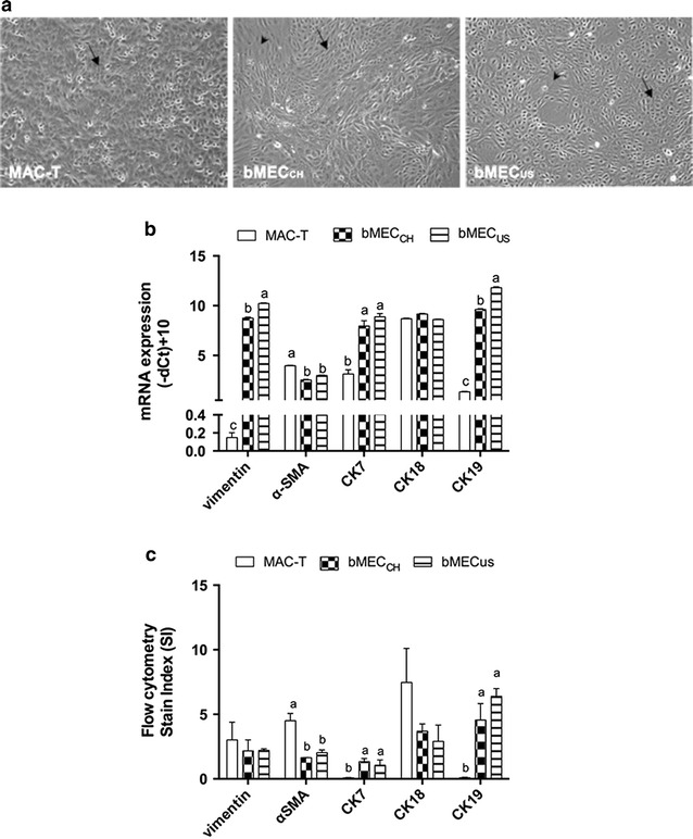
a. Morphology of bovine mammary cell cultures. The confluent monolayers of the bovine mammary epithelial cells were cultured as described in the text. Black arrows show characteristic cobblestone epithelial cells predominantly present in the monolayer. Black arrowheads depict mesenchymal-like cells. MAC-T: immortalized mammary epithelial cell line, 10× magnification; bMECCH: bovine primary mammary epithelial cells isolated from a Swiss Holstein–Friesian cow at late lactation; 10× magnification; bMECUS: bovine primary mammary epithelial cells isolated from an American Holstein at mid-lactation, 10× magnification. b. The mRNA abundance of the selected markers of mesenchymal-like and epithelial cells in human and bovine mammary cell cultures. The gene expression of vimentin, α-smooth muscle actin (α-SMA), cytokeratin (CK) 7, CK18 and CK19 in MAC-T, bMECCH, and bMECUS was normalized to the mean of beta actin and ubiquitin. Details on the origin of the mammary cell cultures are described in a. Data are shown as mean ± SD of the (−delta Ct) + 10. The values are proportional to the gene expression level. Bars indicate the standard deviation of three independent experiments measured at least in duplicates. Different letters (a–c) indicate significant differences (P < 0.05). c. Protein expression of cell markers using flow cytometry. The protein expression of vimentin, α-SMA, CK7 and CK18 was expressed by using the Stain Index (SI) as described elsewhere [41]. SI = [median fluorescence intensity of positive (MFI) –MFI of negative]/(2 × SD of MFI negative). The MFI was derived from evaluation of flow cytometry data with FLOWJO Data Analysis Software. Data are expressed as mean ± SD of 2–3 independent experiments. Different letters (a, b) indicate significant differences (P < 0.05)
