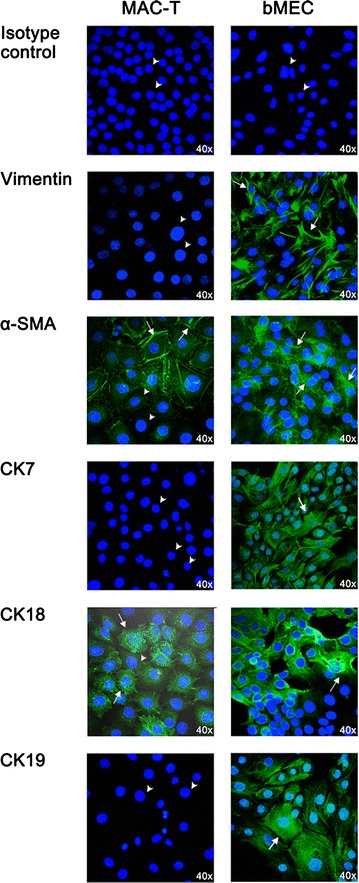
Fig. 4.

Fluorescence microscopy of selected cell markers in bovine mammary cell cultures. The figure shows representative fluorescence microscopy staining of bovine immortalized cell culture (left panel) and primary cell culture (right panel) for vimentin, α-smooth muscle actin (α-SMA), cytokeratin (CK) 7, CK18 and CK19. The negative isotype control IgG1 in each of cell culture is also shown. White arrows show positively stained cells whereas the white arrowheads indicate unstained cells. The fluorescence images were taken with the immunofluorescence microscope Nikon EZ-C1
