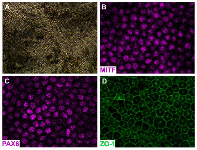
Figure 3. Generation of iPSC derived RPE cells from a control non-LCA individual.
A: Phase micrograph of a confluent monolayer of pigmented iPSC-derived RPE cells. B–D: Immunocytochemical analysis using antibodies targeted against the RPE transcription factors PAX6 (G) and MITF (H), and the tight junction marker ZO1 (I).

