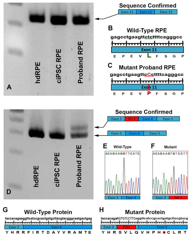
Figure 4.
Investigation of genomic RPE65 variants in human donor eye derived primary RPE and iPSC-derived RPE cells. A: RT-PCR analysis of RPE65 exons 11 to 13 in human control RPE/choroid (hdRPE), human control iPSC-derived RPE cells (ciPSC-RPE) and human LCA iPSC-derived RPE cells (Proband-RPE). A previously identified single heterozygous point mutation detected in exon 11 of DNA isolated from blood (L408P) was confirmed in the proband’s RPE cell transcript. D: RT-PCR analysis of RPE65 exon 3 to 5 in control human donor eye derived primary RPE (hdRPE), human control iPSC-derived RPE cells (ciPSC-RPE), and human LCA iPSC-derived RPE cells (Proband-RPE). An intronic splice site mutation in intervening sequence 3 of the RPE65 gene results in the introduction of a pseudoexon (IVS3 Red) causing a translational frameshift and a premature stop codon. B & C: Schematic diagram depicting wildtype and proband L408P transcript. E & F: Chromatogram depicting of wildtype and proband IVS3-11 transcript. G & H: Schematic diagram depicting wildtype and proband IVS3-11 protein.

