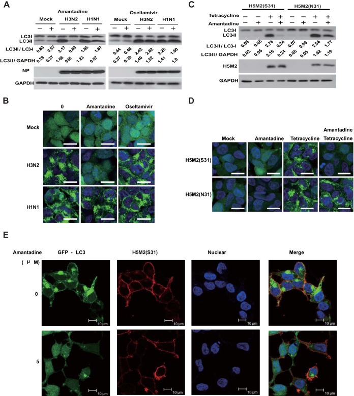
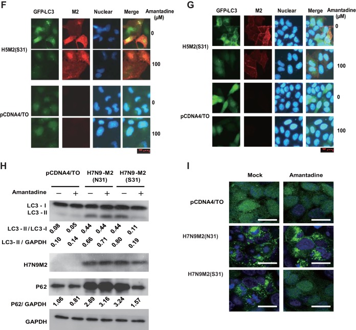
FIG 1.
Inhibition of proton channel activity attenuates influenza A virus M2-induced autophagosome accumulation. (A) HEK293 cells were infected with influenza virus A/Hong Kong/8/68 (H3N2) or A/Wisconsin/33(H1N1) at an MOI of 5. Amantadine (5 μM) or oseltamivir (200 nM) was added at 3 h after infection. Cell were collected at 12 h after infection and subjected to Western blot analysis with the antibodies indicated. NP, nucleoprotein; GAPDH, glyceraldehyde 3-phosphate dehydrogenase. (B) HEK293 cells with stable expression of GFP-LC3 were treated as described for panel A and observed by confocal microscopy. Scale bars, 20 μm. (C) TREx-293 cells carrying tetracycline-inducible amantadine-sensitive H5N1-M2 (S31) or amantadine-resistant H5N1-M2 (N31) were treated with tetracycline (1 μg/ml) with or without amantadine. Cells were collected at 24 h and subjected to Western blot analysis with the antibodies indicated. (D) GFP-LC3-transfected TREx-293 cells carrying tetracycline-inducible H5N1-M2 (S31) or H5N1-M2 (N31) were treated with tetracycline with or without amantadine. Cells were observed 24 h later by confocal microscopy. Scale bars, 20 μm. (E) GFP-LC3-transfected TREx-293 cells carrying tetracycline-inducible amantadine-sensitive H5N1-M2 (S31) were treated with tetracycline with or without amantadine for 24 h. Cells were weakly permeabilized for M2 immunostaining and observed by confocal microscopy. Scale bars, 10 μm. (F, G) MDCK (F) or MCF-7 (G) cells stably expressing GFP-LC3 were transfected, respectively, with plasmids expressing H5M2(S31). Amantadine was added at 6 h after transfection. Cells were collected at 24 h and subjected to immunofluorescence staining with anti-M2 antibodies and a DyLight 549-labeled goat anti-mouse secondary antibody. The cells were observed by fluorescence microscopy. Scale bars, 20 μm. (H) HEK293 cells were transfected with plasmid pcDNA4, pcDNA4-H7N9-M2 (S31), or pcDNA4-H7N9-M2 (N31) and then treated with or without amantadine at 6 h after transfection. Cell were collected at 24 h after amantadine treatment and subjected to Western blot analysis with the antibodies indicated. (I) HEK293 cells with stable expression of GFP-LC3 were transfected with plasmid pcDNA4, pcDNA4-H7N9-M2 (S31), or pcDNA4-H7N9-M2 (N31) and then treated with amantadine at 6 h after transfection. Cells were observed by confocal microscopy at 24 h after amantadine treatment. Scale bars, 20 μm. Representative data from one of three separate experiments are shown. The relative LC3-II/LC3-I, LC3-II/GADPH, and P62/GADPH ratios were analyzed with ImageJ (National Institutes of Health) in the same Western blot assay. The anti-M2 mouse serum was generated in our lab, the anti-influenza A virus nucleoprotein antibody was Abcam Ab139361, the anti-LC3-II antibody was Sigma-Aldrich L7543, and the anti-P62 antibody was Sigma-Aldrich P0067.


