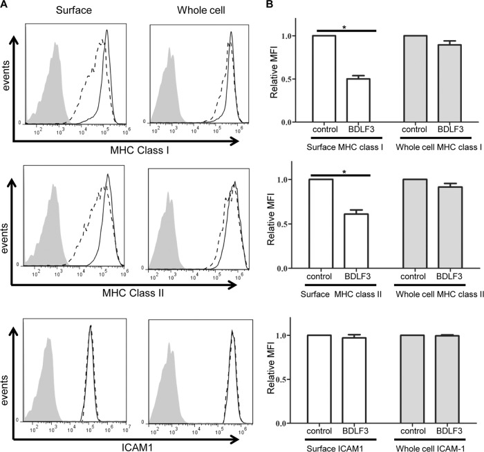
FIG 5.
BDLF3 induces a more dramatic reduction in surface MHC class I and II than in whole-cell MHC levels. (A) MJS cells were transiently transfected with the control-GFP or BDLF3-GFP plasmid. At 24 h posttransfection, two-color flow cytometry was used to measure the levels of surface MHC class I (upper left), MHC class II (middle left), and ICAM1 (lower left) in the viable GFP+ populations of control-GFP-transfected cells (solid lines) and BDLF3-GFP transfected cells (dashed lines). The gray histograms denote background staining obtained with an isotype control antibody. In parallel, these GFP+ transfected MJS cells were analyzed for whole-cell levels of MHC class I (upper right), MHC class II (middle right), and ICAM1 (lower right), using intracellular staining of fixed and permeabilized cells. The results are representative of repeated experiments. (B) Relative mean fluorescence intensities (MFI) of MHC class I, MHC class II, and ICAM1 were calculated for BDLF3-GFP+ cells compared to control GFP+ cells. Results are combined data from three independent experiments. White bars represent surface staining, and gray bars represent whole-cell staining. Differences that reached significance (P < 0.05) by Student's paired t test are denoted by asterisks.

