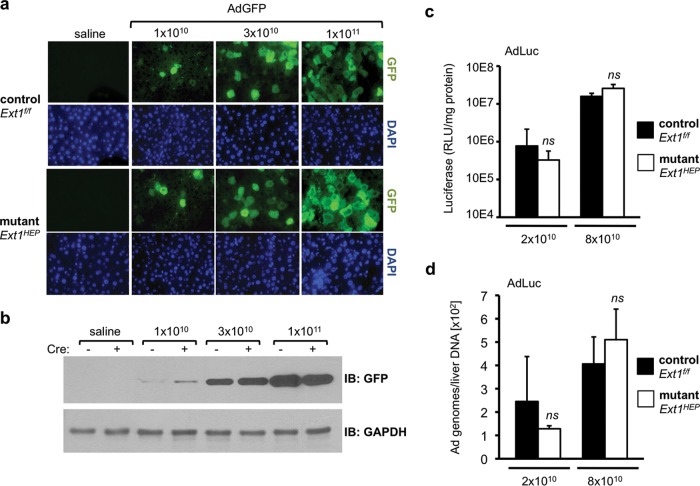
FIG 4.
Adenovirus transduction in Ext1f/f control and Ext1HEP mutant mice. (a) GFP transgene expression in livers of Ext1f/f control or mutant Ext1HEP mice 3 days after intravenous injection with increasing doses (1 × 10¹⁰, 3 × 10¹⁰, or 1 × 10¹¹VP/mouse) of an Ad5 vector expressing GFP. GFP content was visualized by immunofluorescence staining of paraffin-embedded liver sections. Corresponding fields stained with DAPI are displayed to visualize nuclei. (b) Liver extracts were analyzed by immunoblotting for GFP and GAPDH. Liver extracts from two representative mice per group are shown. (c and d) Liver transduction using a low dose (2 × 10¹⁰ VP/mouse) and a high dose (8 × 10¹⁰ VP/mouse) of AdLuc, an Ad5 vector encoding firefly luciferase. Luciferase transgene expression (c) and adenovirus vector genomes (d) in Ext1f/f control and Ext1HEP mutant mice were analyzed 3 days after intravenous injection. Ad5 genomes were detected in liver by quantitative PCR. Values are means ± SD. Differences in either transgene expression or vector genomes between control and mutant mice were compared by Student's t test. ns, not significant (P > 0.05); n = 4 mice per group.

