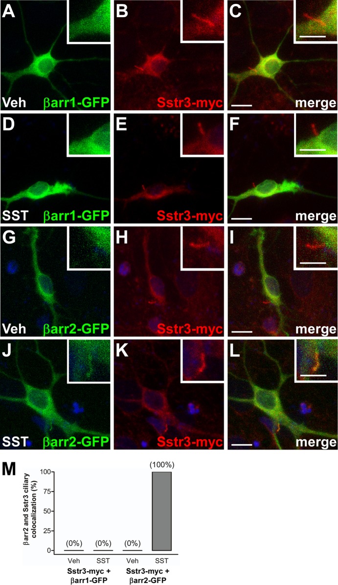
FIG 3.
Somatostatin-mediated localization of β-arrestin 2 to neuronal cilia. (A to L) Representative images of hippocampal neurons from WT mice, after 7 days in culture, transfected with βarr1 fused to GFP (βarr1-GFP; green) (A to F) or βarr2 fused to GFP (βarr2-GFP; green) and myc-tagged Sstr3 (Sstr3-myc; red) (G to L). Neurons were treated with vehicle (Veh) (A to C and G to I) or 10 μM SST (D to F and J to L) for 20 min, fixed, and labeled with an antibody to myc. Sstr3-myc is targeted to cilia. βarr1-GFP was not detected in cilia in vehicle- or SST-treated neurons. βarr2-GFP localized to cilia in SST-treated neurons. (Insets) Higher-magnification images of the cilia. Nuclei were stained with DRAQ5. Bars, 10 μm (main images) and 5 μm (insets). (M) Percentages of Sstr3-positive neuronal cilia that were positive for βarr1-GFP or βarr2-GFP after 20 min of vehicle or 10 μM SST treatment. In vehicle-treated cultures. βarr1-GFP localized to 0% of Sstr3-positive cilia (n = 25) and βarr2-GFP localized to 0% of Sstr3-positive cilia (n = 25). In SST-treated cultures βarr1-GFP localized to 0% of Sstr3-positive cilia (n = 25) and βarr2-GFP localized to 100% of Sstr3-positive cilia (n = 25).

