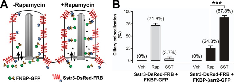
FIG 6.
The ciliary localization of FKBP-βarr2-GFP is greater in response to somatostatin treatment than rapamycin treatment. (A) Schematic of rapamycin-induced dimerization in the ciliary compartment. FKBP-GFP diffuses into and out of the cilium. In the absence of rapamycin, the level of FKBP-GFP in the cilium is low. In the presence of rapamycin, FKBP dimerizes with FRB fused to Sstr3, which is enriched in the ciliary membrane, thereby trapping FKBP-GFP in the ciliary compartment. (B) IMCD cells were transfected with Sstr3 fused to DsRed and FRB (Sstr3-DsRed-FRB) and FKBP fused to GFP (FKBP-GFP) or βarr2-GFP (FKBP-βarr2-GFP). Cells were treated with DMSO (vehicle [Veh]), 100 nM rapamycin (Rap), or 10 μM SST for 10 min and fixed, and the GFP ciliary localization was quantified. In DMSO-treated cultures, FKBP-GFP and FKBP-βarr2-GFP localized to 0% of Sstr3-positive cilia (n = 33 to 38). In rapamycin-treated cultures, FKBP-GFP localized to 71.6% ± 4.8% of Sstr3-positive cilia (n = 41) and FKBP-βarr2-GFP localized to 24.8% ± 5.3% of Sstr3-positive cilia (n = 44). In SST-treated cultures, FKBP-GFP localized to 3.7% ± 3.7% of Sstr3-positive cilia (n = 33) and FKBP-βarr2-GFP localized to 87.8% ± 4% of Sstr3-positive cilia (n = 45). The percentage of FKBP-βarr2-GFP ciliary colocalization was significantly greater after SST treatment than rapamycin treatment. Values are expressed as the mean ± SEM. ***, significantly different results (P < 0.001).

