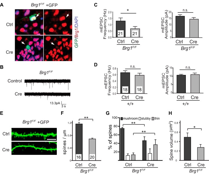
FIG 1.
Deleting Brg1 in hippocampal neurons impairs synapse/dendritic spine formation and maturation. (A) Organotypic hippocampal slice cultures from P6 wild-type or Brg1F/F mice were biolistically transfected with Cre-expressing plasmids or empty vector controls together with GFP. Immunostaining showed Brg1 deletion in Cre-expressing GFP-labeled cells but not in the control cells 5 days after transfection (arrows). (B to D) In the organotypic hippocampal slice culture system, synaptic function was measured using whole-cell patch clamp recordings of Cre-transfected CA1 pyramidal neurons or neighboring untransfected neurons. Representative traces of mEPSCs are shown (B). Average mEPSC frequency and mEPSC amplitude from Cre-expressing (n = 21) or untransfected (n = 21) Brg1F/F CA1 neurons (C) and from Cre-expressing (n = 18) or untransfected (n = 18) wild-type neurons (D) are shown. (E to H) Organotypic hippocampal slice cultures from P6 Brg1F/F mice were biolistically transfected with GFP- and Cre-expressing plasmids or with a vector control. Representative pictures of dendritic spines of CA1 pyramidal neurons are shown in panel E (scale bar, 5 μm). Average dendritic spine densities (F), classifications (G), and spine volumes (H) of control (n = 16) and Brg1-deleted (n = 20) CA1 neurons are shown. Values in the graphs represent averages plus standard errors. **, P < 0.01; *, P < 0.05 (Student's t test). DAPI, 4′,6′-diamidino-2-phenylindole.

