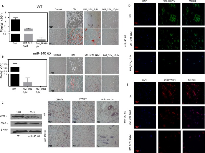
FIG 1.
SFN treatment inhibits preadipocyte differentiation in primary SVF. (A) Sulforaphane treatment (5 or 10 μM) resulted in a dramatic decrease in differentiation, as evidenced by a decrease in lipid droplet accumulation. Primary SVF following differentiation (the 9th day after stimulation with d-biotin, dexamethasone, insulin, and IBMX cocktail) was stained with Oil Red O. (B) Primary SVF obtained from miR-140 KO mice showed a decrease in differentiation in both the presence and absence of SFN (5 or 10 μM). Oil Red O staining indicated that the knockout of miR-140 resulted in a decrease in differentiation, and SFN treatment (5 or 10 μM) blocked adipocyte differentiation. (C) Protein analysis of differentiated WT and miR-140 KO adipocytes for adipocyte markers CEBP/α and PPARγ and IHC imaging of mammary tissue sections from WT and miR-140 KO mice for adipocyte markers, CEBP/α, PPARγ, and adiponectin. Brown precipitate was developed using an avidin-biotin peroxidase substrate kit (Vector Laboratories, CA). Nuclei were counterstained with hematoxylin. The images were captured using a Nikon-Ti microscope. (D and E) Immunofluorescence imaging of differentiated adipocytes. The SVF from miR-140 KO was allowed to differentiate in an adipogenic cocktail with or without SFN (5 or 10 μM) for 10 days. After differentiation, cells were fixed in 4% paraformaldehyde (PFA) and analyzed for the expression levels of mature adipocyte markers (CEBP/α and PPARγ). The cells were stained with anti-rabbit Alexa 488- or 555-conjugated secondary antibody, and nuclei were counterstained with DAPI. Shown are representative images from experiments done in triplicate. The scale bars represent 100 μm. The data represent means and SE.

