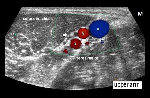
Figure 3.

Transverse scan at the upper arm (just distal to the axilla) shows normal appearance of the right ulnar nerve (dotted ellipse; Cross-sectional area=5 mm2). The nerve comprises of multiple hypoechoic dot-like nerve fascicles (honeycomb appearance on a transverse scan). The arrow points to nerve fascicles of the median nerve that are not oriented transversely on the present scan. [a=high bifurcation of the brachial artery, v=basilic vein, M=medial aspect].
