
Figure 4.
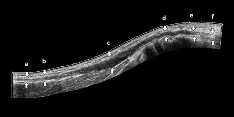
Panoramic view of the right ulnar nerve from the arm to the upper forearm through the cubital tunnel (d) demonstrates normal segments of the nerve (a, f), transition zones (b, e), region of fusiform thickening (c, d). The nerve is maximally thickened (c) proximal to the segment within the cubital tunnel (d).