FIG 1.

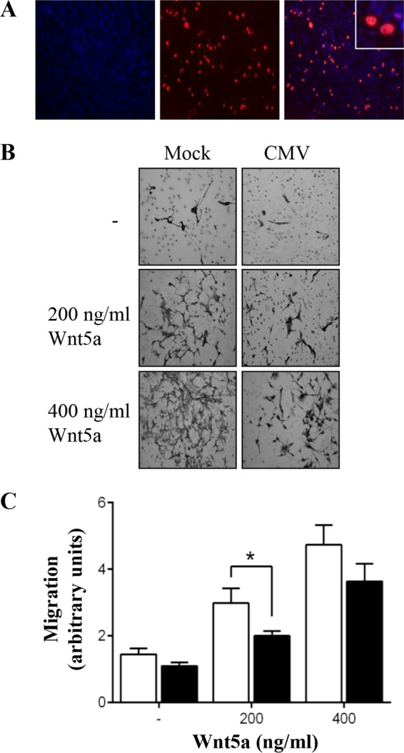
CMV infection inhibits Wnt5a-induced migration of SGHPL-4 trophoblasts. Human trophoblasts were infected with CMV (■) at an MOI of 1 PFU/cell or mock infected (□). (A) CMV-infected cells were analyzed by immunofluorescence at 3 days p.i. Representative images of CMV IE/E protein (red) and DAPI (blue) are shown. (B) Cell migration was analyzed using Transwell assays, with or without addition of recombinant Wnt5a protein. Images are representative of four independent experiments. (C) Average migration, combined from four independent experiments, was determined by capturing five images (20 fields per treatment) from each well and measuring the intensity using ImageJ software (means + SEMs; *, P < 0.05).
