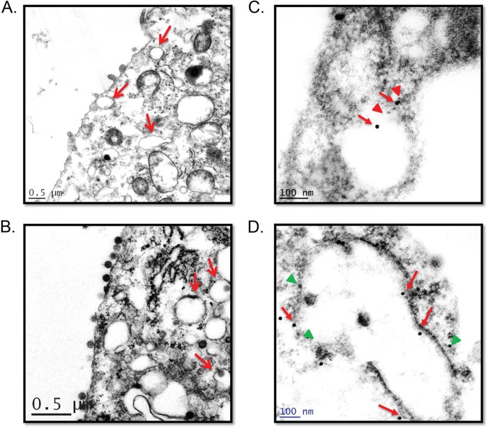
FIG 1.
SFTS virus NSs induces the formation of endosome-like structures. Ultrastructure analyses of SFTS virus NSs-expressing cells (A) and SFTS virus-infected cells (B) show cytoplasmic structures reminiscent of early endosomes (arrows) in ultrathin sections. Immunogold staining of distinct ultrathin sections shows cytoplasmic structures positive for SFTS virus NSs and Rab5 (C) or SFTS virus NSs and LC3B (D). A solid red arrows indicate SFTS virus NSs in panels C and D, while a red triangle (in panel C) and a green triangle (in panel D) indicate the detection of Rab5 and LC3B, respectively. Ultrathin sections of mock-infected cells were also labeled, as indicated above (not shown), to ensure the specificity of antibody. Representatives images are shown.

