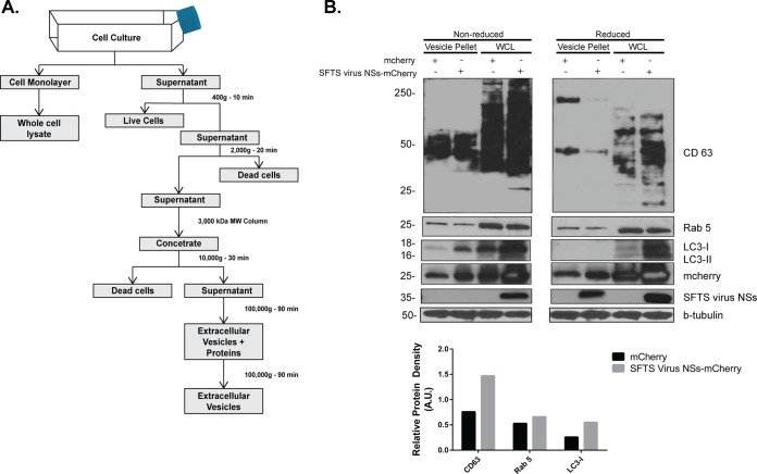
FIG 3.
Isolation and characterization of SFTS virus NSs-positive secreted extracellular vesicles. (A) Schematic representation of the protocol for the isolation of secreted extracellular vesicles by ultracentrifugation. (B) Supernatants from cell lines expressing the mCherry and SFTS virus NSs-mCherry proteins were collected, and isolation of extracellular microvesicles was performed as indicated in panel A. The final pellet was resuspended in lysis buffer, sonicated, resolved by SDS-PAGE electrophoresis, transferred to a PVDF membrane, and blotted for SFTS virus NSs, LC3B, and common markers for microvesicles such as Rab5, β-tubulin, and CD63 (core protein, 26 kDa; glycosylated protein, 30 to 60 kDa). The cell monolayer was used to generate the whole-cell lysate (WCL) and assayed for the detection of the proteins indicated above. Densitometry analysis of CD63, LC3-I, and Rab 5 present in extracellular vesicles isolated from mCherry or SFTS virus NSs-mCherry expressing cells was also conducted. The band signal intensity of each protein was normalized to the signal intensity of β-tubulin and expressed as arbitrary units (A.U.). Signal intensities were obtained by using ImageJ software.

