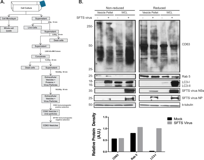
FIG 4.
Characterization of extracellular microvesicles secreted during SFTS virus infection. HeLa cells were mock infected or infected with SFTS virus for 72 h. (A) Supernatant was collected, and isolation of extracellular microvesicles was performed as indicated. (B) The final pellet was resuspended in lysis buffer, sonicated, resolved by SDS-PAGE electrophoresis, transferred to a PVDF membrane, and blotted for SFTS virus NSs, SFTS virus NP, and LC3B, in addition to common markers for microvesicles such as Rab5, CD63, and β-tubulin. The cell monolayer was used to generate the WCL and assayed for the detection of proteins indicated above. Densitometry analysis of CD63, LC3-I, and Rab 5 present in extracellular vesicles isolated from mock infected or SFTS virus-infected cells was carried out. The band signal intensity of each protein was normalized to the signal intensity of β-tubulin and expressed as arbitrary units (A.U.). Signal intensities were obtained by using ImageJ software.

