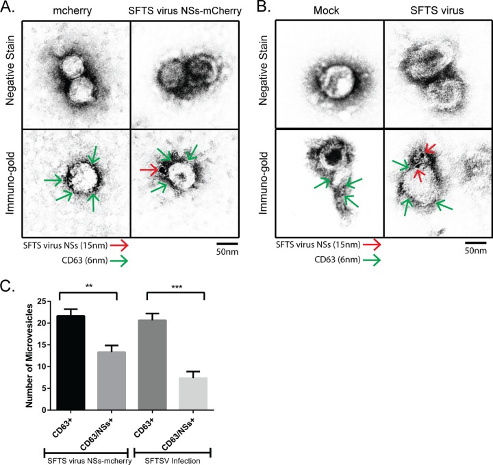
FIG 5.
Isolated extracellular vesicles are positive for SFTS virus NSs and the exosomal marker CD63. Supernatant was collected from HeLa cells stably expressing mCherry or SFTS virus NSs-mCherry (A) and from HeLa cells mock-infected or infected with SFTS virus (B), and isolation of extracellular microvesicles was performed as depicted in Fig. 3A. The final pellet was resuspended in molecular-grade water. The sample was adsorbed onto Ni grids and negatively stained with 2% aqueous uranyl acetate. Additional grids were incubated with primary antibodies against SFTS virus NSs (rabbit) and CD63 (mouse) and then secondary goat anti-mouse IgG couple to 6-nm colloidal gold and goat anti-rabbit IgG coupled to 15-nm colloidal gold antibodies and then negatively stained with 2% aqueous uranyl acetate. SFTS virus NSs is indicated by red arrows, and CD63 is indicated by green arrows. (C) The number of CD63-positive and CD63/SFTS virus NSs-positive extracellular vesicles was quantified based on immunogold labeling observed from 10 fields of view. A total of 22 vesicles were observed in each experiment, of which an average of 14 were positive for both SFTS virus NSs and CD63. All electron microscopy experiments were repeated three times. Results are expressed as means plus the standard errors of the mean (SEM). Asterisks specify statistically significant difference (P < 0.05) between the indicated groups.

